What is the tip of your finger made up of?
The Tendon that allows the tip of your finger to straighten, is called the Extensor Digitorum Communis. This tendon anchors below the nail bed and can be cut or ripped from the bone. The Extensor tendon is a cable that runs from the forearm into your finger and inserts onto the Distal Phalanx (Medical name of the last bone in your finger tip)
Symptoms of a Mallet finger
A Mallet finger is not usually very disabling. Patients do however often tell me that their finger hooks on their pockets when trying to put their hands in their pockets.

Avulsion fracture (Piece of bone ripped out)
- Severe Bruising after 5 – 10 minutes
- Swelling and bleeding visible under the skin (Blue)
- Constant pain irrespective of movement
- Loss of finger movement
- No changes in sensation (normal feeling on the skin)
Tendon rupture
- Bruising (mild)
- Minimal swelling
- No pain when not moving the finger
- Unable to straighten your finger (pain is tolerable)
- No changes in sensation (normal feeling on the skin)
Don’t
Must Do
Makes it worse
Diagnosis
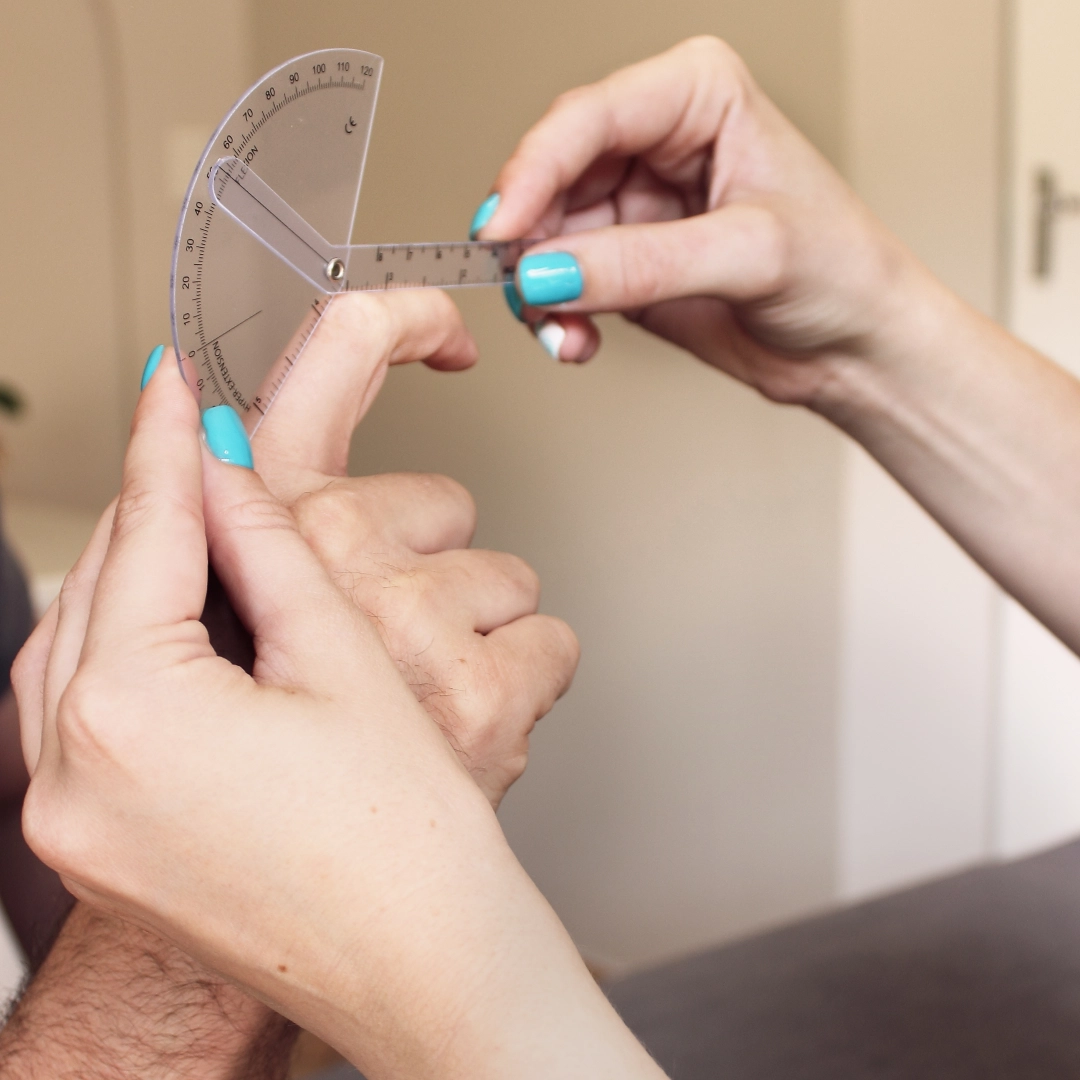
We pride ourselves in our experience with regards to testing the different types of problems that can cause a Mallet finger. Our specialists are able to detect the extent of the damage. We mainly test the three components of a Mallet finger. The Extensor tendon (tendon), the integrity of the Distal Phalanx (bone) and the Distal interphalangeal joint (joint).
We develop a certain dexterity to identify a fracture but in some cases an Ultrasound or X-rays may help with making a more accurate diagnosis.
Why is my finger staying bent?
The Extensor tendon is not able to reconnect by itself, thus staying in this bent position. Splinting the tip of your finger in a straight position is the only way to allow the two loose ends to attach.
If a piece of bone is ripped out, it may cause the bone to heal in the wrong position, or not at all. If a separation of more than 3mm between the bone fragments are present the chances of reattachment is very poor.
A big problem we see with a Mallet finger these days:
When delaying treatment the tendon may never reattach or bone fragments will heal in an awkward position that can permanently deform your finger.
Delaying treatment for more than 2 months, might lead to a Swan Neck deformity. This is when the middle joint of the finger hyperextends and the tip of the finger stays bent. You will have considerable difficulty to use your hand, specifically to grab, hold, pull or grasp objects.
Generic braces may just make the situation worse if you don’t know what you are dealing with. Rather consult with us, to give you a clear diagnosis and prognosis.
We are usually confronted with patients that wait too long before seeking treatment. We cannot stress this enough, because this can significantly change the time to recover your finger back to its normal function.
Treatment for Mallet finger:
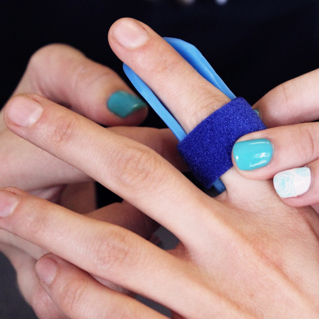
Splinting – A custom splint designed and tailored for your finger, to allow optimal healing and ensure the tendon or bone re-attaches.
Passive Exercises – to maintain PIPJ movement
Active exercises – to regain and strengthen the EDC muscle.
Tendon gliding techniques – to ensure the tendon moves like it should
Scar tissue mobilizations – to guide and provent and abnormal scar tissue adhesions.
Sensory retraining – to reconditions the nerve endings in your finger.
Healing & Recovery Time
Tendon ruptures and stable fractures start with an 8 week immobilisation period, followed by a 4 week rehabilitation protocol. Total time to full recovery: 3 months.
Displaced fractures and surgery start with immobilisation for 4 – 6 weeks using a K-wire, thereafter splinted for 4 – 6 weeks, followed by a 4 weeks rehabilitation protocol. Total recovery time: 4 months.
Medical management
Pain medication may ease some of the pain, but will do nothing to solve the problem.
Surgery of Mallet finger
K-wires are thin wires that the surgeon threads through the bones (like when making a necklace with beads) to keep the Distal Phalangeal joint stable or even inserted through the fractures fragment to join the bone.
The surgeon will insert the K-wire under general anaesthesia and remove it 4 – 6 weeks later. Getting the surgery done is only half-way to recovery. Many patients assume after 6 weeks they will have made a full recovery but a comprehensive rehabilitation program will still follow the surgery.
About our hand therapy services
Other Injuries
Our Partner
Well Health Pro is a medical group of professionals that treat a wide range of muscle, joint, tendon & nerve problems.

